A Large Vessel Found Within the Pericardial Sac
Largest artery in the body. What structure is anterior to the heart.

Heart Anatomy Anatomy And Physiology I
The central portion of the mass was hypointense on.
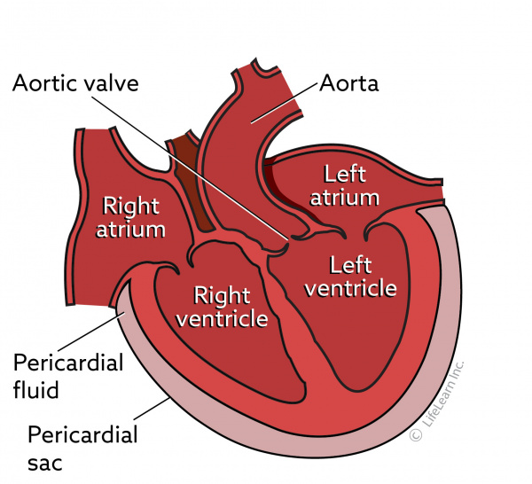
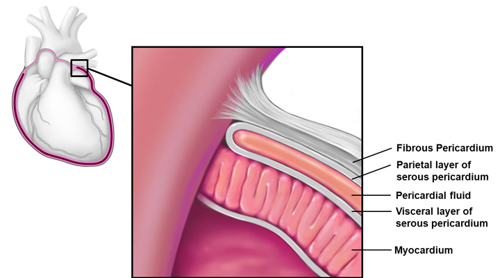
. The portion of the aorta supplying the upper extremities neck and head. The large vessel that branches into the subclavian and carotid. The pericardial sac has two layers a serous layer and a fibrous layer.
A large vessel found within the pericardial sac. What is a large vessel found within the pericardial sac. The pericardium is a double-walled sac containing the heart and the roots of the great vessels.
In addition the patient underwent echocardiography with normal ejection fraction and valve function and with the presence of small pericardial effusion in the posterior pericardial sac. The large vessel that branches into the right subclavian artery and the right common carotid artery. The portion of the aorta supplying the upper extremities neck and head.
Slipping her hand under the heart apex she slid her fingers upward and to the right within the sac until they were stopped by the cul-de-sac formed by the pericardial reflection near the base of the heart. Anterior to the heart is. The portion of the aorta supplying the upper extremities neck head.
The large vessel that branches into the right subclavian artery and the right common carotid artery. The pericardial sac has two layers a serous layer and a fibrous layer. The pericardium is a double-walled sac containing the heart and the roots of the great vessels.
The chest CT showed large pleural effusion on the left and minimal fluid on the right and minimal pericardial effusion Figure Figure2. On histopathological evaluation there were multifocal to coalescing large aggregates of fibrin within pericardium sac and epicardium with a marked diffuse infiltration of neutrophils Figure 1C as well as mesothelial proliferation and blood vessels in the pericardial sac. A large vessel found within the pericardial sac.
The pericardial sac has two layers a serous visceral layer and a fibrous parietal layer. It encloses the pericardial cavity which contains pericardial fluid. Magnetic Resonance imaging MRI of the heart was requested which revealed a large heterogeneous mass within the pericardial sac adjacent to the free wall of the LA and the mid-basal portion of the LV lateral wall measuring 93 x 80 x 43 cm in diameter and multiple septa inside Figure 1.
It encloses the pericardial cavity which contains pericardial fluid. Since the pericardium was torn inferiorly the surgeon began to explore for fragments in the pericardial sac. The pericardium acts as mechanical protection for the heart and big vessels and a lubrication to reduce friction between the heart and the surrounding structures.
It encloses the pericardial cavity which contains pericardial fluid. Pericardium The heart is covered by pericardium - also called pericardial sac which is a double-walled sac containing the heart and the roots of the great vessels. The portion of the aorta supplying the upper extremities neck and head.
It encloses the pericardial cavity which contains pericardial fluid. Slipping her hand under the heart apex she slid her fingers upward and to the right within the sac until they were stopped by the cul-de-sac formed by the pericardial reflection near the base of the heart. Since the pericardium was torn inferiorly the surgeon began to explore for fragments in the pericardial sac.
A very important role in all aspects of pericardial functions is played by mesothelial cells. The pericardial sac has two layers a serous layer and a fibrous layer. A large vessel found within the pericardial sac.
Multifocal and coalescent areas of coagulative necrosis surrounded by intact and degenerate neutrophils. A large vessel found within the pericardial sac. The large vessel that branches into the right subclavian artery the right common carotid artery.
The pericardium is a double-walled sac containing the heart and the roots of the great vessels. The pericardial sac or pericardium is a dual-walled sac that contains the heart and the roots of the great vessels vena cavae pulmonary arteries pulmonary veins aorta brachiocephalic.

Nurse Teaching Cardiac Nursing Critical Care Nursing

Pin On Phlebotomy Anatomy
Pericarditis
Cardiac Tamponade Definition Causes Symptoms And Treatment

Pin On Icu Nursing

Heart Pericardium Great Vessels Thoracic Nerves Flashcards Quizlet

Pericardial Cavity An Overview Sciencedirect Topics

Illustration Picture Of Anatomical Structures Pericardial Sac

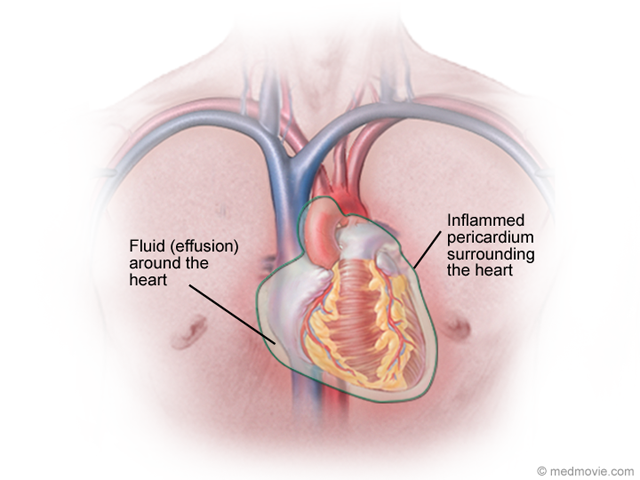
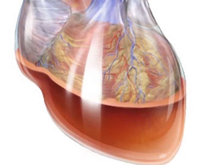
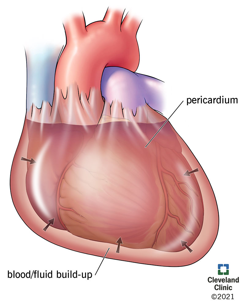
Pericardial Effusion Causes Symptoms And Treatment
![]()
Pericardium Anatomy Of Fibrous And Serous Layers Kenhub
Cardiac Tamponade Causes Symptoms Treatment

8 Large Pericardial Effusion Bright Area With A Typical Swinging Download Scientific Diagram

Pericardial Cavity An Overview Sciencedirect Topics

Pericardial Cavity An Overview Sciencedirect Topics
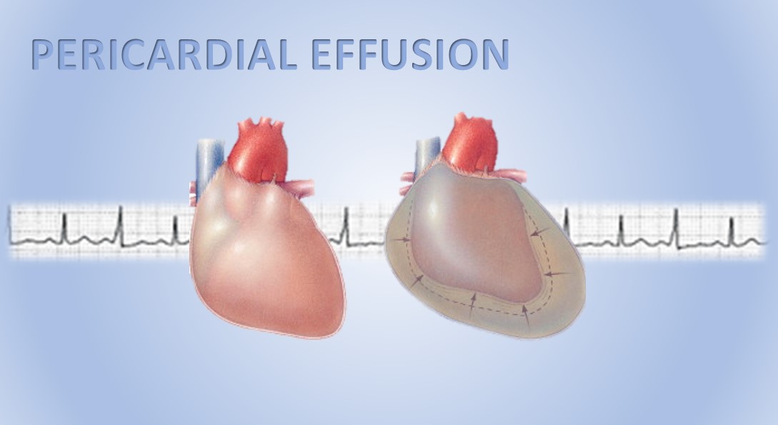
Pericardial Effusion Causes Symptoms And Treatment
Pericardial Effusion In Dogs Vca Animal Hospitals

Pin On Science
Pericardial Disease
![]()
Pericardium Anatomy Of Fibrous And Serous Layers Kenhub


Comments
Post a Comment